Lietuvos chirurgija ISSN 1392–0995 eISSN 1648–9942
2026, vol. 25(3), pp. 295–300 DOI: https://doi.org/10.15388/LietChirur.2026.25(3).8
Superior Mesenteric Artery Syndrome in Children: Diagnostic Challenges and Management Strategies in a Resource-Limited Setting
Neel Aggerwal
Post Graduate Institute of Child Health, Department of Pediatric Surgery, Noida, India
E-mail: aggerwal.pgich@gmail.com
Amit Gupta
Post Graduate Institute of Child Health, Department of Pediatric Surgery, Noida, India
E-mail: amitpedsurgeon@gmail.com
Chandra Vibhash
Post Graduate Institute of Child Health, Department of Pediatric Surgery, Noida, India
E-mail: chandravibhash@yahoo.com
Umesh B. Singh
Post Graduate Institute of Child Health, Department of Pediatric Surgery, Noida, India
E-mail: singhub.2008@gmail.com
Pragya Mishra
Post Graduate Institute of Child Health, Department of Pediatric Surgery, Noida, India
E-mail: tinytots2417@gmail.com
Aditi Solanki
Post Graduate Institute of Child Health, Department of Pediatric Surgery, Noida, India
E-mail: aditisolanki9@gmail.com
Abstract. Background. Superior mesenteric artery (SMA) syndrome is a rare cause of upper gastrointestinal obstruction in children and is often diagnosed late due to nonspecific symptoms and limited awareness. Cases. We report two pediatric cases of SMA syndrome presenting after significant weight loss. The first was a malnourished adolescent with recurrent bilious vomiting in whom diagnosis was delayed due to communication barriers and limited access to imaging; definitive treatment with duodenojejunostomy resulted in excellent recovery. The second child developed acute obstruction following prolonged illness and weight loss; feeding jejunostomy enabled nutritional rehabilitation and complete resolution without the need for definitive bypass surgery. Conclusion. A high index of suspicion is essential for early diagnosis of SMA syndrome in children with upper gastrointestinal obstruction and weight loss. While duodenojejunostomy remains the definitive treatment, feeding jejunostomy may serve as an effective and pragmatic alternative in selected patients, particularly in resource-limited settings.
Keywords: superior mesenteric artery syndrome, children, upper gastrointestinal obstruction, duodenojejunostomy, feeding jejunostomy.
Received: 2025-12-20. Accepted: 2026-01-25.
Copyright © 2026 Neel Aggerwal, Amit Gupta, Chandra Vibhash, Umesh B. Singh, Pragya Mishra, Aditi Solanki. Published by Vilnius University Press. This is an Open Access article distributed under the terms of the Creative Commons Attribution Licence, which permits unrestricted use, distribution, and reproduction in any medium, provided the original author and source are credited.
Introduction
First described by von Rokitansky in 1842, Superior mesentery artery (SMA) syndrome is compression of third part of duodenum between abdominal aorta and SMA [1]. This syndrome arises due to loss of fat pad around duodenum predisposing it to compression, as a result of sudden weight loss or may also be associated with spinal surgery and orthopaedic casting [2]. Symptoms are suggestive of upper gastrointestinal obstruction (UGIO) and range from early satiety, nausea and vomiting and inability to gain weight. Non-specific clinical presentation makes it a diagnosis of exclusion. Past history of major illness causing rapid weight loss strongly indicates the diagnosis. We describe two cases of SMA syndrome highlighting the diagnostic challenges and management strategies in a resource limited setting.
Case 1
The first case was a 15-year-old male child who presented to the emergency department with abdominal pain and bilious vomiting for 15 days. The child was deaf and mute, which made history taking difficult. He was severely underweight (20 kg, <5th percentile) and malnourished. Examination revealed mild to moderate upper abdominal distension with visible bowel loops and peristalsis, but no signs of peritonitis or sepsis. An abdominal radiograph showed a dilated bowel loop with an air–fluid level suggestive of intestinal obstruction.
Initial management included resuscitation with intravenous fluid with nasogastric decompression. Electrolytes were within normal limits, although significant hypoproteinaemia was noted. Subsequently the child tolerated feeds, but at times, he continued to have recurrent bouts of bilious vomiting in same admission. A more detailed history obtained from another close relative later, revealed a history of sudden weight loss during treatment of jaundice 6-years ago following which patient continued to have intermittent symptoms of UGIO and never regained weight. Since the child was deaf and mute, extracting this particular part of history was difficult, as parents were also not much forthcoming.
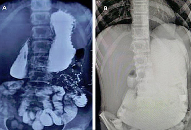
Figure 1. Upper GI contrast study of Case 1 showing grossly dilated stomach and dilated duodenum, DJ flexure in normal location, and delayed gastroduodenal transit.
Upper gastrointestinal endoscopy revealed a grossly dilated stomach and duodenum; however, the endoscope could not be advanced beyond the excessively dilated second part of duodenum. In the absence of an in-house contrast enhanced computed tomography (CECT) facility, a barium meal follow-through was performed, showing a massively dilated stomach (greater curvature descending up to the pelvis), dilated duodenum till proximal third part, delayed gastroduodenal transit and normal duodenojejunal junction position (Figure 1).
At second attempt of endoscopy later, dilatation of the duodenum was seen with external compression resulting in narrowing at the third part of the duodenum. Considering these findings on contrast study and endoscopy along with the history of onset after sudden weight loss due to acute illness, raised suspicion of Superior Mesenteric Artery (SMA) syndrome. Attempts to pass a feeding tube beyond the narrowing were unsuccessful. The patient was placed on conservative management including total parenteral nutrition (TPN) for 2-weeks, arranged through donations. Despite these efforts, his nutritional status did not improve significantly.
After arranging financial support, a CECT scan of the abdomen was done which confirmed the diagnosis of SMA syndrome. Exploratory laparotomy revealed a markedly dilated stomach and proximal duodenum up to the third part, without intraluminal obstruction; a side-to-side duodenojejunostomy was performed between the second part of the duodenum and the jejunum 20 cm distal to the duodenojejunal flexure. The child had an uneventful recovery, achieving full oral intake within 7 days. Over a follow-up period, he gained >5 kg weight after 1 year, gradually increasing to 30 kg over next 2 years and remained completely asymptomatic.
Case 2
Another case was a 11-year-old boy who underwent right hip arthrotomy for septic arthritis which was diagnosed late following a prolonged course of non-specific treatment outside for more than two weeks. On postoperative day (POD) 8, he was referred to the pediatric surgery team with complaints of multiple episodes of retching, bilious vomiting, upper abdominal fullness, and obstipation for the preceding 24 hours, unrelieved by symptomatic treatment. He was emaciated weighing 24 kg and was in hypovolemic shock, with large-volume bilious nasogastric aspirates. Abdominal radiography revealed two prominent air–fluid levels in the right abdomen, suggestive of upper gastrointestinal obstruction. Because of hemodynamic instability and the presence of copious NG aspirates, further investigations were deferred in our resource-limited set-up. After resuscitation, emergency laparotomy showed grossly dilated stomach and proximal third part of the duodenum (D3) with an abrupt transition to a collapsed distal D3; no other obstructive pathology was identified. This background of prolonged course of septic arthritis associated with significant acute weight loss raised suspicion of SMA syndrome. Because the diagnosis remained unconfirmed and the patient was critically ill, a feeding jejunostomy (FJ) was performed to secure reliable enteral access and assist prolonged nutritional rehabilitation if required.
A contrast-enhanced CT scan performed on POD-2 confirmed SMA syndrome, revealing an aortomesenteric angle of 10° and an aortomesenteric distance of 3.4 mm. The patient responded well to jejunostomy feeds, and nasogastric output gradually decreased over 18 days. Refeeding of NG aspirate via the FJ significantly reduced the need for intravenous fluid replacement. The patient transitioned to full oral feeds by day-24 and was discharged with the FJ tube in situ as a precaution. The tube was removed after 2 weeks of asymptomatic period on full oral feeds. On long-term follow-up for over 12 months, he remained asymptomatic and gained approximately 5 kg weight.
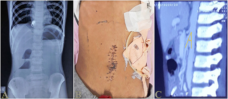
Figure 2. A – pre-operative AXR, B – post-operative clinical image, C – post-operative CECT of Case 2.
Discussion
Superior mesenteric artery (SMA) syndrome is anatomically defined by a reduction of the aortomesenteric angle (normally 20–70°) and aorto-mesenteric distance (10–28 mm) to as low as 6–16° and 2–8 mm, respectively [3]. Although the estimated incidence is 0.013–0.3%, the prevalence in children is even lower [4], making early diagnosis particularly challenging because of non-specific symptoms.
The first case presented to us with acute intestinal obstruction and showed improvement in symptoms following resuscitation. Waxing and waning nature suggested partial UGIO allowed a window for detailed evaluation. Non informative parents and child being deaf and mute, presented as a major block to get a reliable history despite sincere efforts. Diagnosis of SMA relies heavily upon history of sudden weight loss and subsequent appearance of symptoms which is usually elicited via history of patient. But in first case child presented as a usual UGIO and significant history was not retrieved initially due to factors detailed. The second case lost significant weight due to delayed diagnosis of septic arthritis and presented as acute complete UGIO. There are two main causative mechanisms for the pathological anatomy in SMA syndrome: sudden weight loss following debilitating illness or anorexia and spinal corrective surgery (cast syndrome) [4].
The clinical presentation of SMA syndrome is typically non-specific, often overlapping with more common causes of upper gastrointestinal obstruction [1]. Symptoms include post-prandial epigastric pain, bilious vomiting, early satiety, and failure to gain weight. Diagnostic delays are common and may lead to further dehydration, dyselectrolytemia, and worsening malnutrition. In pediatric patients, the condition may also mimic malrotation, duodenal webs, pancreatitis, bezoars, or paraduodenal hernia [3, 4].
Endoscopy may assist by ruling out intrinsic luminal causes [4] and may show external compression at D3, as demonstrated in Case 1. Attempts to place a naso-jejunal tube for enteral feeding may succeed in many patients but were unsuccessful in our first patient due to difficult duodenal negotiation and narrowing, even after two attempts.
Radiological confirmation remains the cornerstone of diagnosis. Upper gastrointestinal contrast studies demonstrate proximal duodenal dilatation, abrupt narrowing at D3, delayed transit, and occasionally relief of obstruction with postural maneuvers–collectively noted in Hines’ criteria [3, 4]. CECT provides direct visualization of duodenal compression along with quantification of the aortomesenteric angle and distance [5]. In Case 1, provisional diagnosis was made based on endoscopy and contrast study due to financial constraints and delayed access to CT scan, whereas in Case 2, the diagnosis was confirmed postoperatively by CECT.
Conservative management is considered first-line and is successful in up to 70% of patients. It includes gastric decompression, small frequent feeds, postural therapy, prokinetics, and nutritional rehabilitation via nasojejunal feeding or total parenteral nutrition (TPN) [4, 6]. In Case 1, prolonged TPN was attempted but financial constraints and inadequate weight gain necessitated definitive surgical management. In Case 2, the child’s acutely unwell condition and uncertain intraoperative diagnosis prompted the choice of a feeding jejunostomy (FJ), which served as a safe intermediate strategy offering enteral access, nutritional restoration, and avoidance of TPN-related complications.
The use of FJ is particularly advantageous in resource-limited settings. In our second case, it allowed rapid improvement in nutritional status, avoided risk of sepsis associated with TPN, facilitated refeeding of naso-gastric aspirates, and ultimately led to complete resolution of obstruction without requiring a more extensive bypass procedure. Moreover, the FJ site remains available for use during future reconstructive surgery if necessary.
Definitive surgical options (open/laparoscopic) include duodenojejunostomy (considered the gold standard with success rates of approximately 90%), gastrojejunostomy, Strong’s procedure (division of the ligament of Treitz), and less commonly, SMA transposition [4, 6, 7]. In Case 1, an open duodenojejunostomy was performed with excellent long-term outcomes. Case 2 improved solely with FJ-assisted feeding, highlighting that in selected patients, restoration of mesenteric fat through enteral nutrition may relieve the obstruction without need for major surgery.
Our two cases highlight the diagnostic challenges of SMA syndrome in children, especially when symptoms are atypical or when comorbid factors–such as communication barriers or acute systemic illness–mask the underlying condition. They also underscore the importance of individualized management. While duodenojejunostomy remains the definitive treatment, FJ may serve as a pragmatic and effective interim or even definitive measure in select pediatric patients, particularly within resource-constrained environments.
Conclusion
Superior mesenteric artery (SMA) syndrome is a rare cause of upper gastrointestinal obstruction in children. A history of sudden weight loss raises high index of suspicion. CECT abdomen confirms the diagnosis. These two cases highlight that there are diagnostic difficulty posed by rarity of the condition, especially when symptoms are atypical or masked by non-reliable history due to communication barriers (like deaf and dumb child), limitations of endoscopy and resource limitations. Feeding jejunostomy may serve as less invasive surgical treatment option avoiding risk associated with major surgical reconstruction.
Author contributions
Neel Aggerwal – conceptualization, investigations, writing original draft.
Amit Gupta – conceptualization, investigations, writing original draft.
Chandra Vibhash – conceptualization, writing original draft.
Umesh B. Singh – visualization, writing review and editing.
Pragya Mishra – investigations, writing review and editing.
Aditi Solanki – writing review and editing.
References
1. Biank V, Werlin S. Superior mesenteric artery syndrome in children: A 20-year experience. J Pediatr Gastroenterol Nutr 2006; 42(5): 522–525.
2. Aldagher A, Almasri R, Mahmoud J. Superior mesenteric artery syndrome in an 8-year-old boy: A case report. J Med Case Rep 2023; 17(1): 343.
3. Hanif M, Siddiqui R, Javed A, Ali M, Farooq O, Fatima M, Farooq H. Superior mesenteric artery syndrome: Delayed diagnosis of a rare clinical entity with a common clinical presentation. Cureus 2022; 14(7): e26729.
4. Oka A, Awoniyi M, Hasegawa N, Yoshida Y, Tobita H, Ishimura N, Ishihara S. Superior mesenteric artery syndrome: Diagnosis and management. World J Clin Cases 2023; 11(15): 3369–3380.
5. Arthurs OJ, Mehta U, Set PA. Nutcracker and SMA syndromes: What is the normal SMA angle in children? Eur J Radiol 2012; 81(8): e854–861.
6. Lee TH, Lee JS, Jo Y, Park KS, Cheon JH, Kim YS, Jang JY, Kang YW. Superior mesenteric artery syndrome: Where do we stand today? J Gastrointest Surg 2012; 16(12): 2203–2211.
7. Mauney CA, Florissi I, Etchill EW, Garcia AV. Laparoscopic duodenojejunostomy for the treatment of pediatric superior mesenteric artery syndrome: A case series. J Pediatr Surg Open 2023; 4: 100065.