Lietuvos chirurgija ISSN 1392–0995 eISSN 1648–9942
2026, vol. 25(3), pp. 313–317 DOI: https://doi.org/10.15388/LietChirur.2026.25(3).11
Giant Breast Lipoma: A Case Report Highlighting an 18-Year Clinical Progression
Saleh AlSuwaydani
Qassim University, College of Medicine, Department of Surgery, Qassim Region, Saudi Arabia
E-mail: s.alsuwaydani@qu.edu.sa
Abstract. Background. Lipoma is the most common benign mesenchymal tumor of adipose tissue, yet its occurrence as a “giant” mass in the breast is rare. Giant breast lipomas are defined by a dimension exceeding 10 cm or a weight greater than 1 000 grams. These lesions pose a significant diagnostic challenge as their massive size and potential for accelerated growth can mimic more aggressive pathologies, such as phyllodes tumors or well-differentiated liposarcomas. Methods. This report describes the clinical presentation, radiological evaluation, and surgical management of a 36-year-old female with no significant comorbidities who presented with a massive right breast lump. The patient reported a notably prolonged 18-year history of the mass, which had entered a phase of accelerated enlargement over the preceding five years, causing physical discomfort and significant mammary asymmetry. Results. Physical examination revealed a soft, mobile, non-tender mass measuring 15x10 cm, without skin changes or chest wall fixation. Computed tomography confirmed a well-circumscribed, fat-density lesion with internal septations and no osseous destruction. The patient underwent complete surgical excision. Histopathological analysis confirmed the diagnosis of a benign giant breast lipoma composed of mature adipocytes with no evidence of cellular atypia or malignancy. Conclusion. Despite an unusually long clinical course and radiological features that may mimic malignancy, giant breast lipomas can be successfully managed through surgical resection. Accurate preoperative imaging and definitive histopathology are essential to ensure appropriate treatment and excellent aesthetic outcomes.
Keywords: breast neoplasms, mesenchymal tumors, giant lipoma, mammary asymmetry, surgical excision.
Received: 2026-04-27. Accepted: 2026-05-17.
Copyright © 2026 Saleh AlSuwaydani. Published by Vilnius University Press. This is an Open Access article distributed under the terms of the Creative Commons Attribution Licence, which permits unrestricted use, distribution, and reproduction in any medium, provided the original author and source are credited.
Introduction
Lipomas are the most common benign mesenchymal tumors composed of mature adipocytes [1]. While they can manifest in any anatomical region containing adipose tissue, the breast is considered a relatively uncommon site for clinically significant presentations, accounting for approximately 16% of all mesenchymal breast neoplasms [2].
Most breast lipomas remain small; however, when they exceed 10 cm in any dimension or weigh more than 1 000 grams, they are clinically classified as “giant” lipomas [2, 3]. These voluminous tumors are rare and frequently present a diagnostic dilemma for surgeons. Due to their massive scale and the potential for phases of accelerated enlargement, giant lipomas can clinically mimic more aggressive conditions, such as phyllodes tumors, breast hamartomas, or primary breast malignancies [2, 3]. Although these lesions are inherently benign, malignant transformation into liposarcomas is exceedingly rare [1, 4]. Nevertheless, the presence of internal septations, traversing vessels, or rapid growth as observed in the later stages of long-standing cases necessitates rigorous radiological and histopathological confirmation to exclude well-differentiated liposarcoma [1, 2].
Surgical intervention is the definitive management strategy for giant breast lipomas, primarily indicated to alleviate physical discomfort, correct significant mammary asymmetry, and address the psychological distress associated with cosmetic deformity [1, 3]. Accurate preoperative imaging is essential to delineate the lesion from the underlying pectoralis fascia and to prevent overtreatment [2]. We report a unique case of a giant breast lipoma in a 36-year-old female, characterized by a notably prolonged 18-year clinical course and a subsequent 5-year phase of accelerated growth.
Case presentation
A 36-year-old female with no significant comorbidities presented for evaluation of a chronic, painless swelling of the breast. The patient reported a total duration of 18 years, during which the lesion remained largely indolent; however, the clinical course was marked by a phase of accelerated growth over the preceding five years. While the mass was predominantly asymptomatic, its progressive enlargement led to physical discomfort and significant mammary asymmetry, prompting the patient to seek surgical consultation for cosmetic correction.
On physical examination, the patient was hemodynamically stable. Localized assessment revealed a soft, well-circumscribed mass measuring about 15x10x6 cm along medial quadrants of right breast (Figure 1A). The lump was neither adherent to the underlying pectoral fascia nor had any associated skin changes, such as tethering, ulceration, or nipple retraction. There was no palpable axillary lymphadenopathy.

Figure 1.
A. Preoperative clinical photograph demonstrating a massive, protuberant mass in the right breast with significant mammary asymmetry.
B. CT scan chest – lung/bone window.
C. CT scan chest – soft tissue/mediastinal window. Axial view. CT sections showing a large, well-circumscribed, hypodense subcutaneous mass in the right anterior chest wall with fat attenuation. Sagittal view. Reconstructions demonstrating the craniocaudal extent of the lesion, showing significant anterior displacement of the skin and breast parenchyma. Coronal view. Images illustrating the transverse dimensions and the relationship of the mass to the midline, revealing internal septations and traversing vessels without chest wall invasion.
Computed tomography (CT) of the chest revealed a well-defined, large, right upper anterior chest wall subcutaneous lesion extending toward the midline, measuring 4.1x14x11.3 cm (Figure 1B–C). The mass was predominantly of fat density, though it featured internal septations with enhancement reaching a maximum thickness of 2.2 mm. Additionally, few calcific foci were noted in the anterior aspect, and prominent vessels were seen traversing the lesion. A subtle smudge of contrast was observed along the mid-aspect; however, no appreciable soft tissue density, chest wall invasion, or underlying osseous destruction was evident. Given the lesion’s size and internal characteristics, the primary differential diagnosis included a giant anterior chest wall lipoma versus a well-differentiated liposarcoma.
The patient was counselled, and informed consent for elective excision of the right breast lump under general anesthesia was secured. A transverse incision was made, revealing a well-encapsulated, lobulated, yellowish mass in the subcutaneous plane, consistent with lipomatous tissue (Figure 2A). The mass was being carefully dissected from the surrounding breast tissue and pectoralis fascia. The excised specimen was a large, lobulated mass measuring approximately 16 cm in its maximum dimension (Figure 2B) and 720 g in weight. The surface was smooth and encapsulated, with a characteristic bright yellow, greasy appearance. Microscopic examination demonstrated a classic benign lipoma composed of mature adipocytes (Figure 2C). The cells were uniform in size with eccentric, flattened nuclei and large, clear cytoplasmic vacuoles. No evidence of cellular atypia, lipoblasts, or malignancy was noted. The postoperative phase was uneventful, and the surgical site healed with a well-approximated transverse scar (Figure 2D). Significant cosmetic improvement and restoration of breast symmetry were evident. The patient was highly satisfied with the outcome.
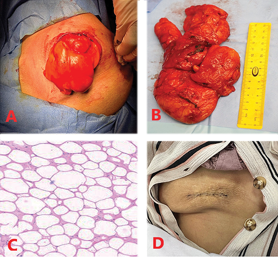
Figure 2. Surgical excision and histopathology:
A. Intraoperative view. Dissection of a well-encapsulated, lobulated yellowish mass through an elliptical incision.
B. Gross specimen. The excised 16 cm specimen demonstrating a smooth, encapsulated surface and characteristic fatty appearance.
C. Histopathology (H&E). Microscopic sections showing uniform, mature adipocytes with no evidence of atypia or malignancy.
D. Postoperative outcome. Healed surgical scar with significant improvement in breast symmetry and contour.
Discussion
Lipomas are the most prevalent form of mesenchymal neoplasms, yet their occurrence within breast tissue is relatively infrequent, with an estimated incidence of 2.1% [1, 5]. While the majority of these lesions are small and managed conservatively, a “giant breast lipoma” is defined by a maximum dimension exceeding 10 cm or a weight greater than 1 000 grams [2, 3, 6]. Some researchers classify lipomas as giant if their maximum dimensions are greater than 5 cm and/or weigh more than 500 g [7]. Our case is particularly notable for its 18-year clinical duration, which aligns with the characteristically slow, indolent progression of benign lipomatous tumors before they reach massive proportions [1, 5].
The etiology of the accelerated growth phase observed in this patient over the last five years remains complex. While lipomas are generally stable, sudden volumetric increases in long-standing lesions can be triggered by hormonal shifts, trauma, or metabolic changes [2]. In the context of a giant mass, this rapid expansion often necessitates a rigorous differential diagnosis to exclude well-differentiated liposarcoma (WDL) or phyllodes tumors [1, 3]. Radiologically, Computed Tomography (CT) and Magnetic Resonance Imaging (MRI) are indispensable for characterization. Features such as thick internal septations (>2 mm), nodular non-adipose components, and prominent internal vascularity are traditionally viewed as red flags for malignancy [5, 6]. However, as demonstrated in this case, these findings can also be present in giant benign lipomas due to repetitive minor trauma or internal fat necrosis [1, 5].
Historically, surgical excision remains the gold standard for management. While some literature explores liposuction-assisted removal to minimize scarring, this approach is often contraindicated for giant lesions due to the risk of incomplete capsule removal and a higher incidence of local recurrence [2, 8]. For this patient, a formal surgical excision was prioritized to ensure complete removal of the encapsulated mass and to achieve immediate restoration of mammary symmetry.
The anatomical localization of breast lipomas is a critical factor in both surgical planning and symptomatic presentation. Most mammary lipomas, including the one described in this case, are subcutaneous, residing within the superficial fatty layer of the breast [2, 3]. However, deeper variants such as subpectoral or retromammary lipomas can occur, which often present with more pronounced displacement of the breast parenchyma and may require a more extensive dissection of the pectoral fascia to ensure complete margin clearance [9, 10]. In our patient, while the giant mass caused significant anterior bulging, its subcutaneous position and the presence of a distinct cleavage plane facilitated a safe plane of dissection without necessitating muscle resection.
Despite being a relatively straightforward procedure, the excision of giant breast lipomas carries a specific set of postoperative risks. The most common complications include seroma and hematoma formation, arising from the significant “dead space” created by the removal of a large-volume mass [2]. In our case, careful intraoperative hemostasis and potentially the use of a suction drain are essential strategies to mitigate these risks. Other documented complications include paresthesia due to the stretching or injury of cutaneous nerves, skin flap necrosis if the dissection is too superficial, and persistent mammary asymmetry or ptosis following the removal of the volumetric support provided by the tumor [2]. For the latter, a secondary mastopexy is sometimes indicated to achieve optimal symmetry once the tissues have settled [6].
Histopathological confirmation is the definitive diagnostic step [6, 7]. The presence of mature adipocytes without cellular atypia or lipoblasts confirmed the benign nature of the lesion, despite the preoperative imaging features concerning preoperative imaging features. This case underscores that even with a notably prolonged history and suspicious radiological signs, a giant breast lipoma can be successfully managed through complete surgical resection with excellent functional and aesthetic outcomes.
Declarations
Conflict of interest. The author declares no potential conflicts of interest with respect to the research, authorship, and/or publication of this article.
Funding. This case report received no specific grant from any funding agency in the public, commercial, or not-for-profit sectors.
Patient consent. Written informed consent was obtained from the patient for the publication of this case report and any accompanying images. A copy of the written consent is available for review by the Editor-in-Chief of this journal.
Declaration of generative AI and AI-assisted technologies in the writing process. During the preparation of this manuscript, the author used generative AI technologies to assist in the linguistic refinement, structural organization, and formatting of the clinical data. After using these tools, the author reviewed and edited the content as needed and takes full responsibility for the clinical accuracy, integrity of the data, and the final intellectual content of the article. The author confirms that no AI tools were used for the generation of clinical data, primary diagnosis, or medical decision-making.
References
1. Alnefaie SM. A giant, recurrent, subpectoral breast lipoma: Case report. Int Surg J 2024; 11: 433–435. DOI: 10.18203/2349-2902.isj20240326.
2. Hassouni FEL, Benchine A, Marrakchi S, Rouas L, Fathi K, Youssfi M. Giant breast lipoma: A case report. Int J Surg Case Rep 2024; 120: 109787. DOI: 10.1016/j.ijscr.2024.109787.
3. Busbaih Z, Almohammed Saleh AA, AlMaghlouth MK, Albeladi AM, Alali T, AlGhadeer MS, Odeh A. Giant breast lipoma: A case report. Cureus 2022; 14(2): e22304. DOI: 10.7759/cureus.22304.
4. Alsalamah R. Giant breast lipoma presenting with right breast triple size the left breast associated with pain and limitation of activity: A case report. Int J Biomed 2025; 15(1): 220–221. DOI: 10.21103/Article15(1) _CR4.
5. Li YF, Lv MH, Chen LF, Wu YF. Giant lipoma of the breast: A case report and review of the literature. Clin Breast Cancer 2011; 11(6): 420–422. DOI: 10.1016/j.clbc.2011.04.005.
6. Yarso KY, Bellynda M, Ismail EA, Kamil MR. Wisepatern mastopexy for natural breast ptosis symmetrization after giant lipoma excision: Case report. Int J Surg Case Rep 2023; 109: 108532. DOI: 10.1016/j.ijscr.2023.108532.
7. Hawary MB, Cardoso E, Mahmud S, Hassanain J. Giant breast tumors. Ann Saudi Med 1999; 19(2): 174–176. DOI: 10.5144/0256-4947.1999.174.
8. Sharma S, Rayamajhi A, Chapagain A, Shrestha JM. A 5 kg giant breast lipoma in a 40-year-old woman: A case report. Ann Med Surg (Lond) 2024; 86(2): 1229–1233. DOI: 10.1097/MS9.0000000000001683.
9. Su A, Margolies L. Large subpectoral lipoma on screening mammography. J Radiol Case Rep 2017; 11(9): 22–27. DOI: 10.3941/jrcr.v11i9.3098.
10. Simango S, Ramdial PK, Madaree A. Subpectoral post-traumatic lipoma. Br J Plast Surg 2000; 53(7): 627–629. DOI: 10.1054/bjps.2000.3396.