Lietuvos chirurgija ISSN 1392–0995 eISSN 1648–9942
2024, vol. 23(2), pp. 120–126 DOI: https://doi.org/10.15388/LietChirur.2024.23(2).6
Correction of Constricted Ear – Case Report
Ljubinka Damjanoska Krstikj
University Surgery Clinic “St. Naum Ohridski”, Skopje, Republic of North Macedonia
E-mail: ljubinka_damjanoskakrstik@yahoo.com
Ana Rajkovska Kimovska
University Surgery Clinic “St. Naum Ohridski”, Skopje, Republic of North Macedonia
E-mail: ana_rajkovska@yahoo.com
Roza Krsteska
Faculty of Medical Sciences, Goce Delchev University, Shtip, Republic of North Macedonia
E-mail: roza.krsteska@ugd.edu.mk
Mare Stevkovska
University Surgery Clinic “St. Naum Ohridski”, Skopje, Republic of North Macedonia
E-mail: mare_st@yahoo.com
Abstract. Introduction. Congenital deformities of the outer ear are common; reported incidence are from 1:6000 to 1:6830 newborns. Multitude of corrective procedures are described in the literature. Purpose of the case study. The adequate surgical treatment of the congenital constricted ear remains a challenge. Selected tailor made approach should be employed on case by case basis, thus, in this study we present our case of moderate constricted ear and the used surgical procedures done under general anesthesia, as well as the outcome of the treatment. Case report. A 6 year old girl presented in pediatric and plastic surgery department with complaint of right ear deformity from birth and additional psychological effects such as increased difficulty in social integration and lack of self- confidence. On clinical examination, we considered as constricted ear grade IIB by Tanzel. Two separate procedures under general anesthesia were done. In the first procedure, advancement of the helix and otoplasty was performed; an effective expansion, of about 1 cm in the length of the pinna was obtained. In the second procedure, about 0.5 cm expansion of the helix was performed, and also the breadth of the pinna. Conclusion. In our case, combination of Mustardé suture, helix advancement and helix cartilage graft in moderate constricted ear were a useful surgical option, producing aesthetically good results in a simple and effective way.
Keywords: congenital malformation, constricted ear, surgical treatment.
Received: 2024-01-10. Accepted: 2024-02-28.
Copyright © 2024 Ljubinka Damjanoska Krstikj, Ana Rajkovska Kimovska, Roza Krsteska, Mare Stevkovska. Published by Vilnius University Press. This is an Open Access article distributed under the terms of the Creative Commons Attribution Licence, which permits unrestricted use, distribution, and reproduction in any medium, provided the original author and source are credited.
Introduction
Abnormal development or deformities of the ear anatomy can cause a range of complications, from cosmetic issues to hearing and development problems. An estimated 5 to 45 percent of children are born with some sort of congenital ear deformity. Approximately 5% of the population suffers from some degree of ear prominence. The incidence of outer ear malformations has been reported at 1:6000 newborns to 1:6830 newborns [1, 2]. Some ear deformities are temporary. If the deformity was caused by abnormal positioning in the uterus or during birth, it may resolve as the child grows, the ear unfolds and takes on a more normal form.
Other ear deformities will need medical intervention – either nonsurgical or surgical – to correct the ear anomaly. Because it’s unknown which ear deformities will correct on their own and which will not, it’s important to discuss treatment options as early as possible.
Prominent ears, constricted ear, cup ear are the most well-known facial disfigurements influencing youngsters.
Because outer ear deformities are present from birth, a number of factors may contribute to their development:
• Environment. Congenital ear deformities can occur when a developing baby is exposed to certain conditions in the uterus. Prenatal exposure to particular drugs, including isotretinoin (Accutane, for example), thalidomide, and alcohol have been linked to the development of outer ear deformities.
• Fetus positioning. In some cases, deformities arise due to how a baby is positioned in the uterus or during birth. For instance, a reduction in blood supply to the outer ear can lead to abnormal development.
The constricted ear is an auricular deformity produced by a deficiency in the circumference of the helical rim. While the exact cause of constricted ear is unknown, all types of the deformity occur due to some tissue deficiency (cartilage or skin), resulting in the helix (ear rim cartilage) not developing adequately. Constricted ear is not simply a misshapen ear, but rather the ear is missing portions of skin and/or cartilage. In severe cases, which involve a more abnormally shaped ear in which normal auricle components are absent, constricted ear presents as a form of microtia, a condition characterized by parts of the external ear being malformed, smaller than normal or missing completely. Microtia, however, often involves hearing loss and inner ear defects, while patients with constricted ear usually have no hearing difficulties. Symptoms of constricted ear can include: ddecreased ear size, lop deformity (folded forward and down), low ear position and protrusion.
Cup ear refers to the deformity of the ear where the top rim of the ear is folded, tight, or wrinkled. The condition of a cup ear can vary from mild to severe. Where in the mild condition only the rim of the upper ear is folded, in the severe form the helix cartilage and scapha of the ear is squeezed forming a roll.
The classification and corrective methods for constricted ears continue to be controversial [3]. The constricted ear deformities described by Weerda [4] following Tanzer [5] are classified as follows:
Type I (slight deformity) affects only the helix. A slight cap-shaped projection of the helix hangs over the scapha; the crus inferius and the helicis are normally present. The longitudinal axis of the pinna is slightly shortened. Often there is concomitant prominence of the ear.
In Type II deformity, the helix, the anthelix with its crura and the scapha are affected.
• Type IIa (slight to moderate deformity) shows a hood-like overhang of the helix accompanied by flattening or absence of the crus superius antihelices and a pronounced crus inferius antihelices. The shortening of the longitudinal axis of the pinna is greater. The ear is often prominent. Straightening of the pinna rarely reveals skin defects.
• In Type IIb (moderate to strong deformity), the hood-shaped helix overhang and the shortening of the longitudinal axis are more marked. The ear is decreased in width, particularly at the upper part. The crura of the antihelix and the antihelix itself are flattened or absent. The pinna is prominent, and straightening usually reveals insufficient skin.
Type III (severe deformity) shows marked underdevelopment of the upper pinna, extreme overhanging of the superior auricular components and considerable deficits in height and width of the ear. There is often dystopia, showing low and anterior positioning, and EAC stenosis is frequently present, occasionally EAC atresia.
More than 200 techniques have been used for the surgical correction of prominent ears, referring that no single “best” method exists and that techniques and modifications will continue to appear [6–8].
Constricted ears undergoing surgical correction, most of them were performed with:
• helical expansion through auricular or costal cartilage graft;
• mustardé-type mattress sutures, and tumbling cartilage flap;
• external molding using Vaseline gauze rolls was implemented on every case to assist reshaping the scapha;
• a triangular superficial temporal fascial flap was elevated to prevent the reoccurrence of lidding (covering) in some cases.
Non-surgical treatment for constricted ear, lop ear, and cup ear is available when sought early, within the first few weeks of life. There is correction system which is highly successful for correcting many ear deformities, including constricted ears, when utilized shortly after birth. There is correction system, four-piece molding system which painlessly reshapes the ears while the cartilage is still soft and malleable and can be placed during a short office visit and remains on the ear for approximately four to six weeks. Some benefits of this system include: non-surgical, permanent, and painless method of correcting ear deformities, very high 90% success rate and fast results (usually in 2 months).
Purpose of the case study
Adequate surgical treatment of the congenital constricted ear remains a challenge, despite the multitude of corrective procedures described in the literature. Selected tailor made approach considering best practices should be employed on case by case basis, thus we present our case of moderate constricted ear. We used two stage technique, done under general anesthesia. In this study we present the technique and the outcomes of it.
Case report
6 year old girl had constricted ear grade IIB by Tanzel, on her right ear: decreased ear size, lop deformity (folded forward and down) and protrusion (Figure 1).


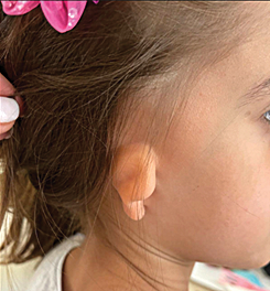
Figure 1. Initial status
Two stage operation was performed:
First stage. Antihelix reconstruction (Mustardee suture) and crus helix advancement.
Second stage. Helix expansion with split concha cartilage flap.
For the first operation while the patient was under general anesthesia an incision was made through the skin and cartilage around the crus of the helix. Crus helix was repositioned superiorly and retro aproximatly about 10 mm. On that way the pinna was unfold and reopened.
Posteriorly on the pinna was made an incision and was made three Mustardee suture for corection of otopostasis of the outer ear.
The retroauricular skin incision was performed 8–10 mm below and parallel to the helical rim. The skin above the cartilage was mobilised caudally up to the mastoid and cranially to the helical rim. In order to prevent postoperative skin distortions, the mobilization should not be extended beyond the helical rim. The perichondrium, which ensures adequate nutrition of the cartilage, and the auricular cartilage itself remain intact. The new antihelices fold was punctured with needles from ventrally. Subsequently, the mattress sutures made of nonabsorbable, transparent Prolen 4.0, were placed at the corresponding markings, using a retroauricular access through the auricular cartilage and the perichondrium, without penetrating the ventral skin. The knots of the mattress sutures were everted towards the inside to prevent protrusion through the skin.
At the end of the operation an effective expansion, of about 1 cm in the length of the pinna was obtained (Figure 2).
A  B
B 
A. Before; B. After
Figure 2. Status after the first procedure
After 1 year, a second operation was performed and a free cartilaginous graft was taken from the concha and interposed in the region of the upper pole of the helix, thereby elongating the helix and the upper pole of the auricle. Excess retroauricular skin was excised.
The procedure was done under general anesthesia, and its starts as the procedure for otoplasty. The retroauricular skin incision was performed 8–10 mm below and parallel to the helical rim. The skin above the cartilage was mobilized caudally up to the mastoid and cranially to the helical rim and mobilization was beyond the helical rim. Incision of the cartilage was performed about 7 mm below and parallel to the helical rim, around the upper poll of the auriculae, approximately 20 mm. In the middle of the separated helix cartilage was done vertical incision which splits the helix cartilage. In between was interpolate cartilage graft harvested from the concha from the same ear – 10 mm/4 mm, sutured with nonresorbilae 5/0 suture. Skin was overlapped on the new made helix. Excessive retroauriculre skin was excised. Effective expansion, of about 0.5 cm in the breadth of the pinna was obtained.
Auricule was molded with vaseline gauze, and gauzes with rivanoli solution. Ear was fixed with external light compressive elastic bandage.
A  B
B 
C  D
D  E
E 
Figure 3. Selected images from the second procedure
A and B – incision of the cartilage performed about 7 mm below and parallel to the helical rim, around the upper poll of the auriculae, approximately 20 mm. C and D – interpolate cartilage graft harvested from the concha from the same ear – 10 mm/4 mm, sutured with nonresorbilae 5/0 suture. E – molded auricule with vaseline gauze, and gauzes with rivanoli solution.
Discussion
According to some authors, if the correction starts early, right after birth, can give excellent results and thus the child will not be subjected to surgery and to the socio-psihological pressures from the environment [9, 10]. If this period is missed, then the operation should be done as soon as possible, before starting school, in order to save the child from the pressures of the environment, especially from her friends at school.
There are several techniques [11] some solve only the upper pole, and that is in the case of the first degree of deformity with Z-plasty [12]. With greater bending of the ear shell, there is a lack of cartilage and skin. Cartilage can be taken from the same auricle, from the other, or from a rib and modeled into a new auricle. If there is a lack of skin, then a local skin or fascial flap is taken [13, 14]. It can be done in one or two stages.
In our case, where the auricle was smaller in size, with a protrusion and placed lower in relation to the other ear, there was also a decrease in hearing. We take in considiration that it is a girl and we do not want to leave unnecessery scars on her body, otherwise than the ear shell (for taking cartilage from a rib for example with a scar on the body) we decided to use the material that was available. Due to the presence of protrusion of the auricle, we decided to solve that condition first, but due to the large deformity and short helix, we decided to mobilize the helix at the same time. After the first operation, she had a satisfactory appearance, but what makes the family and the child more happy is the hearing improvement. In the second act, the auricle was opened even more by placing a cartilage graft harvested from the same ear, always taking in a consideration to meake as less as posible scars.
The size of the auricle remained unchanged, but satisfactory opening and repositioning of the auricle was achieved (Figure 4).
A  B
B  C
C 
A. Before; B. After; C. After
Figure 4. Status after the second procedure
Conclusion
Prominent ears are one of the most well-known facial disfigurements influencing youngsters. As for the surgical treatment, despite the multitude of corrective procedures described in the literature, adequate surgical correction of the congenital constricted ear remains a challenge. The maintenance of the shape and size of the reconstructed upper neohelix poses a particular problem. Thus, selected tailor made approach considering best practices should be employed on case by case basis.
For the case presented, two stage procedure, under general anesthesia was done; in first stage antiheliks reconstruction (Mustardee suture) and crus helix advancement and in the second stage helix expansion with split concha cartilage flap, the outcome was successful in aesthetic aspect, improved hearing, an effective expansion, of about 1 cm in the length and about 0.5 cm, in the breadth of the pinna was obtained. And last but not least, as reported by the parents noticeable positive psychological effects in terms of increased self-confidence and improved social integration.
References
1. Brent B. The pediatrician’s role in caring for patients with congenital microtia and atresia. Pediatr Ann 1999; 28(6): 374–383. DOI: 10.3928/0090-4481-19990601-09.
2. Elden LM, Zur KB. Congenital Malformations of the Head and Neck. 1st. New York, NY: Springer, 2014. DOI: 10.1007/978-1-4419-1714-0.
3. Huang X, Ma C, Chang J, Sun P, Wang C, Guo P, Pan B. Classification and surgical strategies of constricted ears in a Chinese specialty clinic: a retrospective study. Aesthetic Plast Surg 2022; 46(5): 2194–2207. DOI: 10.1007/s00266-021-02699-1.
4. Weerda H. Reconstructive facial plastic surgery: a problem-solving mannual. 2nd revised and expanded edition. New York, NY: Thieme Medical Publishers, 2014.
5. Tanzer RC. The constricted (cup and lop) ear. Plast Reconstr Surg 1975; 55(4): 406–415. DOI: 10.1097/00006534-197504000-00003.
6. Pickrell BB, Hughes CD, Maricevich RS. Partial ear defects. Semin Plast Surg 2017; 31(3): 134–140. DOI: 10.1055/s-0037-1603968.
7. Nazarian R, Eshraghi AA. Otoplasty for the protruded ear. Semin Plast Surg 2011; 25(4): 288–294. DOI: 10.1055/s-0031-1288921.
8. Kelley P, Hollier L, Stal S. Otoplasty: evaluation, technique, and review. J Craniofac Surg 2003; 14(5): 643–653. DOI: 10.1097/00001665-200309000-00008.
9. Gulati RD, Faraci N, Butts SC. Neonatal ear molding. Laryngoscope 2021; 131(2): E423–E427. DOI: 10.1002/lary.28842.
10. Kim J, Jo T, Choi J, Kim J, Jeong W. Efficacy of classic ear molding for neonatal ear deformity: case series and literature review. J Clin Med 2022; 11(19): 5751. DOI: 10.3390/jcm11195751.
11. Park C, Park JY. Classification and algorithmic management of constricted ears: a 22-year experience. Plast Reconstr Surg 2016; 137(5): 1523–1538. DOI: 10.1097/PRS.0000000000002120.
12. Duan W, Liu Y. Correction of Tanzer type IIB constricted ears via Z-shaped double V–Y advancement flaps. Ann Plast Surg 2019; 82(3): 284–288. DOI: 10.1097/SAP.0000000000001750.
13. Park C. A new corrective method for the Tanzer’s group IIB constricted ear: helical expansion using a free-floating costal cartilage. Plast Reconstr Surg 2009; 123(4): 1209–1219. DOI: 10.1097/PRS.0b013e31819e2644.
14. Lee JS, Kim JS, Lee JW, Choi KY, Yang JD, Chung HY, Cho BC. Correction of microtia with constriction features using a superficial temporal fascial flap combined with a rib cartilage graft. Arch Plast Surg 2020; 47(4): 317–323. DOI: 10.5999/aps.2018.01165.